S

p

o

r

t

s

H

e

a

l

i

n

g

There are many variations of passages available sure there majority have suffered alteration in some form by inject humour or randomised words which don’t look even slightly believable.

Contact Info

Wellington Hospital, Wellington Knee Unit, Platinum Medical Centre, 15 - 17 Lodge Road, London, NW8 7JA

Knee Passport

Side-to-side stability

The medial and lateral collateral ligaments guide side-to-side motion and protect the knee from collapse. Most MCL injuries heal well without surgery. Understanding the grade and context of your injury guides the right treatment.


ANATOMY

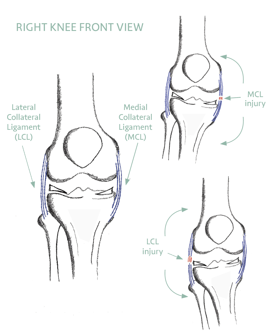
What the Collateral Ligaments Do

The medial collateral ligament (MCL) is a strong band of tissue connecting the femur to the tibia on the inner (medial) side of the knee. It provides stability against forces that push the knee inward — known as valgus forces — and is the most commonly injured knee ligament.

The lateral collateral ligament (LCL) runs along the outer side of the knee, connecting the femur to the fibula. It resists forces pushing the knee outward (varus forces). Isolated LCL injuries are less common, but the LCL can be injured as part of a more complex posterolateral corner injury.


A CLOSER LOOK

Layers, Partners, and Why It Heals

The MCL is not a single cord but two layers working together. The broad superficial layer is the largest structure on the inner side of the knee — roughly 10 to 12 cm long — and is the main restraint against the knee buckling inward. Beneath it, a deep layer blends with and anchors to the medial meniscus, which is why an injury on the inner side of the knee can sometimes involve the meniscus as well.

The MCL does not work alone. Just behind it, the posterior oblique ligament helps control rotation — particularly when the knee is straight. Clinicians describe these structures in terms of primary and secondary restraints: each direction of stress has one main stabiliser plus backups that share the load. The superficial MCL is the primary brake on inward (valgus) force while also providing secondary control of rotation — so even a significant MCL injury rarely leaves the knee without any protection.

There is also a biological reason MCL injuries heal so reliably. Unlike the cruciate ligaments deep inside the joint or the inner rim of the meniscus, the MCL has a generous blood supply — the genicular arteries run directly alongside and through it. Good blood flow delivers the cells and nutrients that repair tissue, which is why most MCL injuries, including many complete tears, mend well with time and rehabilitation rather than surgery.

On the outer side, the LCL is the primary restraint against the knee bowing outward. It rarely tears in isolation; when it does, it is often part of a wider posterolateral corner injury (Destination 9), which is why a careful assessment always looks beyond the ligament itself.

HOW MCL INJURIES HAPPEN

Causes and Grading

MCL injuries typically occur due to a sudden force or impact to the outer side of the knee, causing the ligament to stretch or tear. Common causes include football, skiing, and contact sports. There are three grades:

  • Grade 1 : Minor stretching or micro-tears. Ligament remains intact. Recovery – 1 to 2 weeks
  • Grade 2 : Partial tear. Some instability on testing. Recovery – 3 to 6 weeks
  • Grade 3 : Complete tear. Significant instability. Often heals without surgery. Recovery – 6 to 12 weeks

SYMPTOMS

What You Might Experience

COMMON SYMPTOMS

  • Pain and tenderness on the inner or outer side of the knee
  • Swelling and inflammation around the injured area
  • Stiffness or difficulty bending the knee
  • Instability — a feeling of the knee giving way, particularly during impact or twisting

‘Notice whether your knee falls inward or outward when you climb stairs. This tells you which muscles need attention and where your rehabilitation focus should be.’


TREATMENT

Management — Most Heal Without Surgery

The vast majority of collateral ligament injuries — including many complete Grade 3 MCL tears — heal well without surgery, given the right rehabilitation programme. Time, guided rehabilitation, and a gradual return to load are the key ingredients.


NON-OPERATIVE MANAGEMENT

  • RICE protocol (Rest, Ice, Compression, Elevation) in the acute phase
  • Pain relief and anti-inflammatory medication
  • Bracing or immobilisation to support the knee early
  • Physiotherapy — quadriceps, hip abductor, and proprioception exercises
  • Gradual return to sport

WHEN SURGERY MAY BE INDICATED

  • Persistent instability despite full conservative management
  • Multiple ligament injuries — MCL with ACL or PCL
  • A bony fragment pulled off with the ligament
  • Early return to professional sport required

PREVENTION

Reducing Future Risk

Strengthening the muscles around the knee — particularly the quadriceps and hip abductors — reduces the valgus load on the MCL during activity. Maintaining flexibility through regular stretching, using proper technique in sport, and wearing a protective brace when returning to contact activities can all reduce recurrence risk.


YOUR SELF-CHECK: Stand on one leg and bend your knee slightly. Does your knee drift inward? This is a sign of hip abductor weakness that places extra stress on the MCL. Hip strengthening exercises are as important as knee exercises in MCL rehabilitation.


EVIDENCE BASE

What the Research Shows

REFERENCES — CLINICAL REFERENCES

  1. Phisitkul et al. (Sports Medicine & Arthroscopy Review, 2006) — MCL injuries successfully managed without surgery in the majority of cases across all grades.
  2. LaPrade & Wijdicks (JOSPT, 2012) — The management of injuries to the medial side of the knee: evidence-based treatment algorithm.
  3. Wijdicks et al. (Am J Sports Med, 2010) — Anatomy of the medial part of the knee, detailing the structures involved in MCL injuries.
  4. LaPrade MD, Kennedy MI, Wijdicks CA, LaPrade RF (2015) — Anatomy and Biomechanics of the Medial Side of the Knee. Sports Med Arthrosc Rev. Source for the two-layer MCL, posterior oblique ligament and medial blood supply.
  5. Swinford ST, LaPrade R, Engebretsen L, Cohen M, Safran M (2020) — Biomechanics and physical examination of the posteromedial and posterolateral knee: state of the art. J ISAKOS. Source for the primary/secondary restraint roles.
  6. Amis AA, Gupte CM, Bull AM, Edwards A (2006) — Anatomy of the posterior cruciate ligament and the meniscofemoral ligaments. Knee Surgery, Sports Traumatology, Arthroscopy 14:257–263. Foundational anatomical reference for PCL and related ligament structures by Mr Gupté and colleagues at Imperial.
  7. BASK Research Priorities (Ahmed I, Metcalfe AJ, Baker P, Gupte CM et al., Bone and Joint Journal, 2024) — Identifies ligament and meniscal injury as top research priorities for UK knee surgery, highlighting the clinical importance of optimal management of these injuries.

Ligament Assessment

Get an accurate diagnosis and a personalised rehabilitation plan.