S

p

o

r

t

s

H

e

a

l

i

n

g

There are many variations of passages available sure there majority have suffered alteration in some form by inject humour or randomised words which don’t look even slightly believable.

Contact Info

Wellington Hospital, Wellington Knee Unit, Platinum Medical Centre, 15 - 17 Lodge Road, London, NW8 7JA

Knee Passport

The shock absorber

The menisci are two crescent-shaped structures between the femur and tibia. They distribute load, absorb shock, and protect the bone surfaces. Not all tears need surgery — and understanding the type and context of a tear is essential to making the right decision.


ANATOMY

What is the Meniscus?

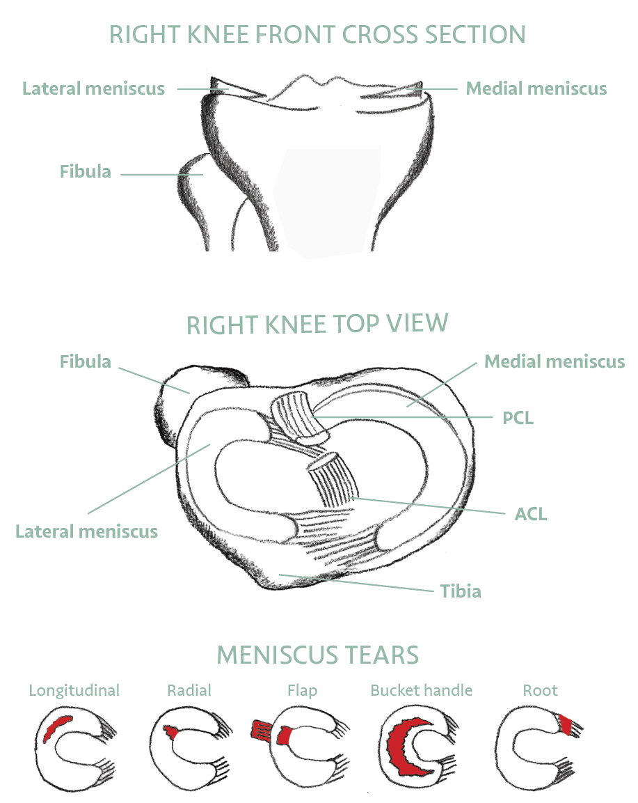
The meniscal cartilages are C-shaped structures that sit between the femur and tibia, helping reduce pressure between the bones and absorbing shock during weight-bearing. There are two menisci in each knee: the medial meniscus on the inner side, and the lateral meniscus on the outer side.

The meniscus is one form of cartilage — known as fibrocartilage, made primarily of Type I collagen. This should be distinguished from the shiny hyaline cartilage (Type II collagen) that lines the bone surfaces and allows low-friction movement. Both are important; both are served by regular movement.

The menisci transmit 50 to 70 percent of load across the knee in extension and up to 85 percent in flexion. They also contribute to joint stability, lubrication, and proprioception — the body’s sense of joint position.


‘When you pivot, turn smoothly and with control. Squat and lunge with good form. The meniscus is stressed most by sudden twisting and deep, uncontrolled bending.’


HOW TEARS HAPPEN

Causes of Meniscal Tears

Meniscal tears occur in two main ways. In younger patients, tears usually involve a significant injury — often twisting and impact — and may be associated with damage to other structures such as the cruciate ligaments. The tear is typically noticed at the time of injury.

In older patients, a gradual weakening and thinning of the meniscal tissue can occur with age, or can be associated with chronic conditions such as diabetes or hypothyroidism. This degenerative process can lead to a small, progressive tear that may not be noticed at the time it develops.


SYMPTOMS

How Do I Know if I Have a Meniscal Tear?

DIAGNOSIS

How is a Meniscal Tear Diagnosed?

Most tears are suspected based on clinical history and examination. An MRI scan is the most reliable way to confirm the diagnosis and characterise the tear — its type, location, and whether it has displaced. X-ray alone cannot diagnose a meniscal tear as the meniscus is soft tissue, not bone.

Movement quality often matters as much as the MRI report. Many people over 40 have degenerative meniscal changes on MRI with no pain at all. The scan must always be interpreted alongside symptoms and clinical examination.

TYPES OF TEAR

Tear Patterns and What They Mean

Different tear patterns have different names, different symptoms, and different treatment implications.


TEAR TYPES

  • Longitudinal — along the length of the meniscus; can sometimes be repaired
  • Radial — across the width; disrupts load distribution significantly
  • Flap — a flap of tissue that can catch and cause symptoms
  • Bucket handle — large displaced tear, often causes locking; frequently requires surgery
  • Root — detachment at the attachment point; significantly affects load distribution
  • Degenerative/horizontal — age-related; common finding, often manageable without surgery

TREATMENT

Should I Have Surgery?

The decision whether to operate depends on many factors including age, activity level, duration of symptoms, whether physiotherapy has been tried, the type of tear, and whether the meniscus has moved out of its normal position.

The British Knee Society (BASK) has published evidence-based guidelines on when meniscal surgery should be considered. Mr Gupté is an executive member of BASK and was involved in developing these guidelines.

Where surgery is indicated, the key question is whether the torn portion can be repaired (stitched back) or needs trimming. This depends on the location of the tear, the blood supply to that zone, the quality of the meniscal tissue, and the pattern of the tear. Tears in the outer vascular zone — the “red zone” — have a better blood supply and are more likely to heal after repair.

Surgery is performed arthroscopically (keyhole) under general anaesthetic, typically taking 15 to 45 minutes. There are usually two small portal scars less than 4 mm in length.


WHEN SURGERY IS USUALLY NOT RECOMMENDED

  • Moderate to severe arthritis already present in the knee
  • Degenerative tears in older patients responding to physiotherapy
  • Significant surgical risk factors — heart disease, blood clots, severe obesity

YOUR SELF-CHECK: Does your knee lock or catch during movement? Locking — the knee freezing and being difficult to fully straighten — is a more urgent symptom than pain alone and usually warrants prompt clinical assessment. Pain with twisting or deep kneeling, without locking, is often manageable with physiotherapy first.


EVIDENCE BASE

What the Research Shows

REFERENCES — CLINICAL REFERENCES

  1. Sihvonen et al. (NEJM, 2013) — Arthroscopic partial meniscectomy was no better than sham surgery for degenerative tears in middle-aged patients without locking.
  2. Katz et al. (NEJM, 2013) — Physical therapy as effective as meniscal surgery for most patients with degenerative tears and mild to moderate OA at 6 months and 2 years.
  3. Abram SGF, Beard DJ, Price AJ & BASK Meniscal Working Group (Bone and Joint Journal, 2019) — The 2018 BASK Arthroscopic Meniscal Surgery Treatment Guideline: a nationally endorsed, evidence-based consensus establishing five clinical presentations and four treatment pathways, from urgent surgery for a locked knee to non-surgical management for degenerative tears with advanced OA. Mr Gupté is a named member of the BASK Meniscal Working Group that produced this guideline.
  4. Hulet C, Pereira H, Peretti G, Denti M (eds.) — Surgery of the Meniscus, ESSKA/Springer, 2016. The definitive European textbook on meniscal surgery; distinguishes traumatic tears (most common: longitudinal, 29% medial, 33% lateral) from degenerative tears and describes the full spectrum of management from repair to replacement. Load transfer: 70% through the lateral meniscus in the lateral compartment; 50% through the medial meniscus medially.
  5. Sherman SL, Chahla J, LaPrade RF, Rodeo SA (eds.) — Knee Arthroscopy and Knee Preservation Surgery, Springer, 2024. State-of-the-art reference covering meniscal repair, ramp lesions (up to 42% prevalence in ACL injuries), meniscus transplant, and inside-out repair techniques; establishes that delayed ACL repair beyond 12 months produces a 3.5× increased odds of medial meniscal tear.
  6. Thorlund et al. (BMJ, 2015) — Systematic review confirming exercise therapy as a first-line option for many meniscal tears.
  7. Gupte CM, Bull AMJ, Thomas RD, Amis AA (2003) — A review of the function and biomechanics of the meniscofemoral ligaments. Arthroscopy 19:161–171. Mr Gupté’s PhD research establishing the anatomy and role of the structures linking the lateral meniscus to the femur.
  8. Gupte CM, Bull AMJ, Thomas RD, Amis AA (2003) — The meniscofemoral ligaments: secondary restraints to the posterior drawer. Journal of Bone and Joint Surgery Br 85(5):765–773. Biomechanical study showing the meniscofemoral ligaments also protect the lateral meniscus and reduce contact pressure.
  9. Amadi HO, Gupte CM, Lie DTT, McDermott ID, Amis AA, Bull AMJ (2008) — A biomechanical study of the meniscofemoral ligaments and their contribution to contact pressure reduction in the knee. Knee Surgery, Sports Traumatology, Arthroscopy 16:1004–1008. Demonstrates that preserving the meniscofemoral ligaments during PCL and meniscal surgery is advisable.
  10. Zhao Y, Coppola A, Karamchandani U, Gupte CM et al. (2024) — Artificial intelligence applied to MRI reliably detects the presence, but not the location, of meniscus tears: a systematic review and meta-analysis. European Radiology. Co-authored with Mr Gupté’s team at Imperial; relevant to MRI interpretation and the limits of AI in meniscal diagnosis.
  11. Tsitsifylla C, Amiras D, Chappell KE, Gupte CM et al. (2026) — In-vivo magic angle MRI imaging reliably identifies collagen fibre orientation in ACL and meniscal tears. British Journal of Surgery. Cutting-edge imaging from the SportsHealing/Imperial research group.
  12. Gupte CM, Smith A, McDermott ID, Bull AMJ, Thomas RD, Amis AA (2002) — Meniscofemoral ligaments revisited: anatomical study, age correlation and clinical implications. Journal of Bone and Joint Surgery Br 84(6):846–851. Comprehensive cadaveric study of the meniscofemoral ligaments in 84 knees, establishing their incidence and age-related changes — the foundational anatomy paper of Mr Gupté’s PhD.
  13. Gupte CM, Smith A, Jamieson N, Bull AMJ, Thomas RD, Amis AA (2002) — Meniscofemoral ligaments: structural and material properties. Journal of Biomechanics 35:1623–1629. First paper to characterise the load-to-failure and elastic properties of the meniscofemoral ligaments, establishing them as capable of transmitting meaningful forces in the knee.
  14. Gupte CM, Bull AMJ, Atkinson HD, Thomas RD, Strachan RK, Amis AA (2006) — Arthroscopic appearances of the meniscofemoral ligaments: introducing the “meniscal tug test”. Knee Surgery, Sports Traumatology, Arthroscopy 14:1259–1265. Describes how to identify the meniscofemoral ligaments during arthroscopy and introduces the clinical tug test — directly relevant to arthroscopic meniscal surgery and repair decision-making.


Meniscus Assessment

Get a clear diagnosis and personalised treatment plan from Mr Gupté and the SportsHealing team.