S

p

o

r

t

s

H

e

a

l

i

n

g

There are many variations of passages available sure there majority have suffered alteration in some form by inject humour or randomised words which don’t look even slightly believable.

Contact Info

Wellington Hospital, Wellington Knee Unit, Platinum Medical Centre, 15 - 17 Lodge Road, London, NW8 7JA

Knee Passport

The glide layer

Chondral cartilage covers the bone surfaces of the knee, allowing near-frictionless movement lubricated by synovial fluid. When it is damaged — through injury, repetitive stress, or a condition called osteochondritis dissecans — the consequences range from persistent pain to early arthritis. Understanding the type and stage of injury is essential to choosing the right treatment.


THE GLIDE LAYER

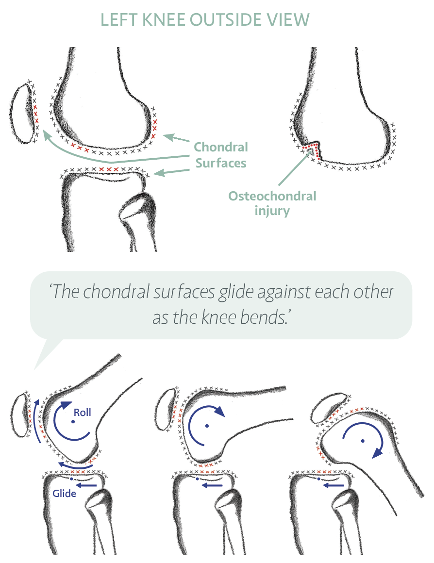
What Chondral Cartilage Is

Articular (hyaline) cartilage is the smooth, glassy tissue covering the ends of the femur, tibia, and the back of the patella. It is between 2 and 4 mm thick and has a coefficient of friction lower than ice on ice. The knee joint produces synovial fluid which both lubricates and nourishes this cartilage — since cartilage has no blood supply of its own, it depends on movement to circulate nutrients.

The chondral cartilage is not to be confused with the meniscus cartilage. The meniscus is fibrocartilage (Type I collagen) acting as a shock absorber between the bones. Chondral or articular cartilage is hyaline cartilage (Type II collagen) acting as the frictionless glide surface on the bone ends. Both types are important; both require protection.

Cartilage does not regenerate easily once damaged. This makes protecting and nourishing it through lifestyle, appropriate exercise, and timely treatment especially important.


‘Meniscus is one type of cartilage. Chondral is another. Regular, gentle movement nourishes both — it circulates the synovial fluid that feeds the cartilage.’


ACUTE INJURY

Osteochondral Injury and Fracture

An osteochondral injury involves damage to both the chondral cartilage surface and the underlying bone. It can occur as a result of a traumatic injury — such as a direct blow, a fall, or the force generated during a knee dislocation — or from repetitive mechanical stress over time.

The symptoms of an osteochondral injury include pain, swelling, stiffness, a sensation of clicking or catching in the joint, and in more severe cases, locking or giving way. These latter symptoms may indicate that a fragment has become loose within the joint.

MRI scan is the most reliable test for confirming and characterising the injury. It shows the size, depth, location, and whether the fragment is stable or at risk of loosening.


NON-OPERATIVE TREATMENT

  • Rest from impact activities
  • Physiotherapy — strength and range of movement
  • Protected weight-bearing if advised
  • Repeat MRI at 3 to 9 months to confirm healing
  • Healing can take 6 to 12 months in milder cases

WHEN SURGERY MAY BE NEEDED

  • Fragment loose in the joint (catching or locking)
  • Defect not healing after 6 months of rest
  • Full-thickness defect with fluid beneath on MRI
  • Lesion in a young, active patient at risk of early arthritis
  • Large defect or one in a high-load area

Surgical options depend on the size, location, and condition of the defect. Most procedures are performed arthroscopically (keyhole), though large defects or those at the kneecap may require a small open incision of 3 to 5 cm.

CHRONIC CONDITION

Osteochondritis Dissecans (OCD)

Osteochondritis dissecans is a condition where a segment of bone and its overlying cartilage loses its blood supply, becomes devitalised, and can eventually detach as a loose body within the joint. It most commonly affects the medial femoral condyle (the inner surface of the thigh bone) in the knee, but can also occur on the patella or the lateral condyle.

OCD most commonly occurs in teenagers and young adults, though it can occur at any age. If the loose fragment detaches and moves within the joint, it can damage the surrounding cartilage and cause early osteoarthritis if not treated.


CAUSES

Why Does OCD Develop?

The exact cause is not fully determined, but the following factors are thought to contribute:


CONTRIBUTING FACTORS

  • Direct injury — a direct blow to the knee surface, for example from a cricket or hockey ball
  • Repetitive stress fracture — high-impact activities repeated consistently over time, including running, jumping, and gymnastics
  • Impaired blood supply — disruption to the local circulation for reasons that are not always apparent
  • Vitamin D deficiency — low bone mineralisation increases susceptibility
  • Rapid skeletal growth — particularly during teenage years
  • Genetic predisposition — a family history increases risk

SYMPTOMS

What You Might Experience

DECISION MAKING

Factors That Determine Treatment for OCD

Whether OCD is managed conservatively or surgically depends on an individual assessment of the following factors:


FACTORS FAVOURING CONSERVATIVE MANAGEMENT

  • Young age — the younger the patient, the greater the potential for spontaneous healing
  • Symptoms present for less than 6 months
  • Stable lesion on MRI — no fluid signal beneath the fragment
  • Small defect size
  • No catching, locking, or giving way

FACTORS FAVOURING SURGERY

  • Swelling, restricted movement, or instability on examination
  • Fluid signal beneath the lesion on MRI — implies instability
  • Symptoms not improving after 6 months of rest
  • Lesion appears loose or displaced on MRI
  • Large defect or high-load location

Repeat MRI scans at 3 to 6 monthly intervals are often the most informative way to monitor whether a lesion is healing or deteriorating before committing to surgery.

If the loose fragment cannot be reattached, or is not in healthy enough condition to fix back, it may need to be removed. The resulting bed in the bone can then be treated with microfracture, a collagen membrane, or an osteochondral transplant depending on size and location.

Once full healing has taken place — which can take 8 to 18 months — the rate of successful return to full sporting activities is approximately 95%. A small proportion of patients experience a recurrent injury or develop a lesion in a different area, but the great majority return to sport with minimal ongoing pain.


RECOVERY

What Rehabilitation Involves After Surgery

Most patients require non-weight-bearing on crutches with a brace for 4 to 8 weeks post-operatively, depending on the procedure. Rehabilitation then progresses through the following stages:


LIFESTYLE SUPPORT

Supporting Chondral Health Every Day

DAILY HABITS THAT PROTECT CARTILAGE

  • Regular low-impact movement — cycling, swimming, walking — circulates synovial fluid
  • Strong muscles absorb joint load and reduce cartilage stress
  • Good nutrition — protein, collagen, Vitamin C, and Vitamin D
  • Healthy body weight to reduce mechanical load on the joint surface
  • Adequate sleep — tissue repair occurs predominantly overnight
  • Avoid sudden large increases in impact training load

WHEN TO SEEK HELP: If knee pain has persisted beyond six weeks, particularly after a direct injury, or if the knee is consistently swollen, catching, or giving way — specialist assessment with an MRI scan is advisable. Early diagnosis of OCD in particular can make the difference between successful conservative management and the need for surgery.


EVIDENCE BASE

Clinical References

REFERENCES — CLINICAL REFERENCES

  1. Buckwalter & Mankin (Journal of Bone and Joint Surgery, 1998) — Articular cartilage repair: fundamental science and current status. Foundational paper on why cartilage fails to self-repair and what that means for treatment strategy.
  2. Husen M, van Egmond N, Custers RJH. In: Sherman et al. (eds.) Knee Arthroscopy and Knee Preservation Surgery (Springer, 2024) — Comprehensive chapter on OCD of the knee. Defines OCD as a focal, idiopathic alteration of subchondral bone with risk for instability and disruption of adjacent cartilage. Peak incidence 12–19 years; male:female ratio approximately 4:1. Most common location: lateral side of the medial femoral condyle (78%). OCD progresses through five distinct pathological stages from subchondral osteopenia to complete fragment displacement. In juvenile OCD, drilling achieves up to 100% healing at 6 weeks to 2 years; in adults, only 25% show healing, making age the most important prognostic factor.
  3. Gross PW, Nagra K, Fabricant PD. In: Sherman et al. (eds.) Knee Arthroscopy and Knee Preservation Surgery (Springer, 2024) — Chapter on osteochondral fracture repair. Lateral femoral condyle and patellofemoral shear injuries require arthroscopic or open fixation to restore articular congruity; treatment depends on fragment size, location, and cartilage viability.
  4. NICE Guidance (2017) — Autologous chondrocyte implantation for treating symptomatic articular cartilage defects of the knee: guidance on when cartilage restoration procedures are appropriate.
  5. Camathias et al. (Knee Surgery, Sports Traumatology, Arthroscopy, 2015) — Systematic review of surgical treatment and outcomes in paediatric OCD: internal fixation with compression screws provides the most reliable healing in unstable lesions with viable bone.

Cartilage and OCD Assessment

Get a specialist diagnosis and tailored treatment plan from Mr Gupté and the SportsHealing team.