IMPORTANT CONTEXT
MRI Findings and Pain Do Not Always Match
Research consistently shows that many people have significant findings on MRI — cartilage changes, meniscal degeneration, small effusions — with no pain at all. A scan report must always be interpreted alongside your symptoms and clinical examination. A diagnosis should never be made from imaging alone.
For suspected arthritis, a weight-bearing X-ray is the correct first investigation. It shows the true joint space under the load the knee actually carries. An MRI taken lying down can appear near-normal even when significant cartilage loss exists under functional load.
Cynthia Gupté (FRCR, MRCS) is a consultant musculoskeletal radiologist and the reporting radiologist for myKneeScan.com — providing specialist-reported knee MRI as part of the SportsHealing diagnostic pathway.
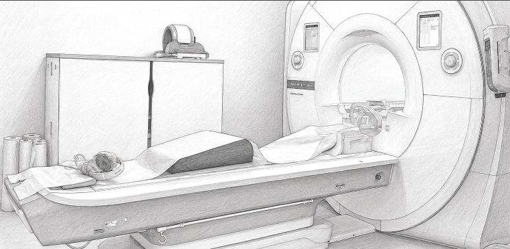
WHAT A SCAN CAN AND CANNOT SEE
MRI Sees Some Things Better Than Others
MRI is not equally good at seeing every structure. It is excellent for some injuries — a torn ACL, for example, is usually clear — but markedly less reliable for others. Injuries to the small, peripheral structures of the posterolateral corner, the popliteus tendon in particular, are easy to under-call on a scan, and almost any injury becomes harder to read weeks or months later, once the acute changes have settled. In those situations a skilled hands-on examination can actually be more sensitive than the scan.
This is the other half of the “interpret in context” message: a scan can show findings that do not cause symptoms, and it can also miss findings that matter. That is precisely why the most accurate diagnosis comes from combining the images with your history and a careful physical examination — never from imaging alone — and why an experienced musculoskeletal radiologist, who knows where these blind spots lie and reads the study alongside your clinical picture, adds so much value.
BEFORE YOUR SCAN: Write down your three main symptoms and when they started. Note whether they are getting better, worse, or staying the same. Bring this to your scan appointment. A radiologist who understands your clinical context will produce a more accurate and useful report.
RESEARCH FROM THE SPORTSHEALING / IMPERIAL TEAM
Mr Gupté’s Publications on MRI and Imaging
REFERENCES — CLINICAL AND RESEARCH REFERENCES
- Zhao Y, Coppola A, Karamchandani U, Gupte CM et al. (2024) — Artificial intelligence applied to magnetic resonance imaging reliably detects the presence, but not the location, of meniscus tears: a systematic review and meta-analysis. European Radiology. Co-authored with Mr Gupté’s team. This is a clinically important finding: AI can flag that a meniscal tear is present, but cannot reliably tell the surgeon which type or where — meaning clinical examination and experienced radiology reporting remain essential.
- Ristic M, Chappell KE, Lanz H, Gupte CM, Amiras D et al. (2024) — First in-vivo magic angle directional imaging using dedicated low-field MRI. Magnetic Resonance in Medicine. This cutting-edge research from Imperial and SportsHealing demonstrates for the first time that MRI can visualise individual collagen fibre bundles in the knee in living patients — potentially transforming how ligament and meniscal injuries are diagnosed.
- Tsitsifylla C, Amiras D, Chappell KE, Gupte CM et al. (2026) — In-vivo magic angle MRI imaging reliably identifies collagen fibre orientation in ACL and meniscal tears. British Journal of Surgery. The clinical application of the above technology, showing that magic angle MRI can improve diagnosis of partial ACL tears and meniscal lesions over conventional MRI.
- NICE Guidelines NG226 (2022) — Weight-bearing X-ray is the appropriate first-line investigation for suspected knee OA. MRI should not be routinely ordered in primary care for OA diagnosis.
- Brinjikji et al. (American Journal of Neuroradiology, 2015) — Systematic review showing high prevalence of imaging findings in asymptomatic individuals — underscoring that all scans must be interpreted in clinical context.
- Swinford ST, LaPrade R, Engebretsen L, Cohen M, Safran M (2020) — Biomechanics and physical examination of the posteromedial and posterolateral knee: state of the art. J ISAKOS. Reports that MRI is less reliable for peripheral and chronic posterolateral injuries, where physical examination can be more sensitive.
- Royal College of Radiologists (2022) — Guidance on appropriate musculoskeletal MRI referral: emphasises clinical correlation and the appropriate use of weight-bearing X-ray as first-line.