S

p

o

r

t

s

H

e

a

l

i

n

g

There are many variations of passages available sure there majority have suffered alteration in some form by inject humour or randomised words which don’t look even slightly believable.

Contact Info

Wellington Hospital, Wellington Knee Unit, Platinum Medical Centre, 15 - 17 Lodge Road, London, NW8 7JA

Knee Passport

The framework of movement

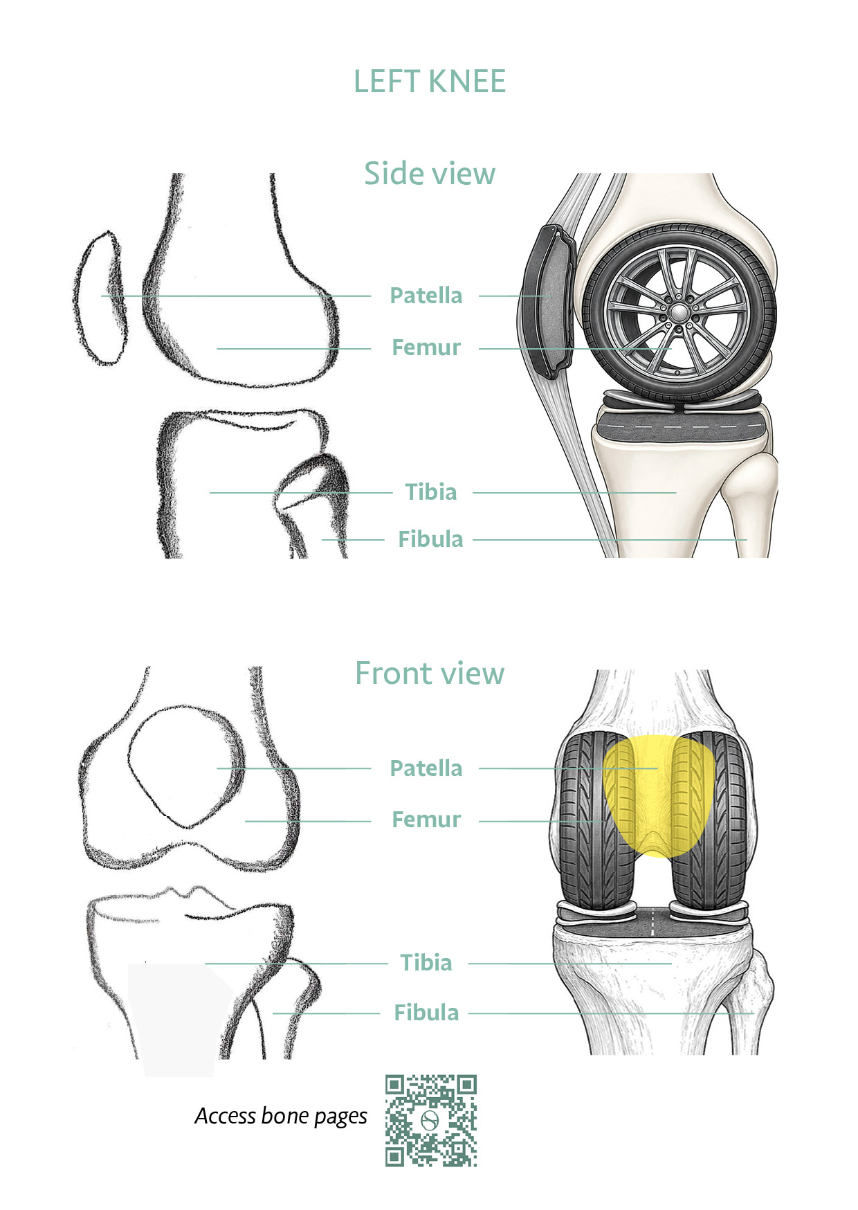
The knee is built from three bones: the femur, tibia, and patella. They form the structure around which every other tissue works — a precisely engineered platform for load, movement, and stability.

ANATOMY

The Three Bones of the Knee


Femur (thigh bone) — The femur ends in two rounded surfaces called condyles that sit on the tibia. These curved surfaces roll and glide during knee movement, allowing a wide range of motion while maintaining stability.

Tibia (shin bone) — The main load-bearing platform. Its upper surface is not perfectly flat: the inner (medial) side forms a shallow dish that cradles the femur, while the outer (lateral) side is gently domed. The whole surface is also tilted very slightly backwards — a feature that helps the knee bend more deeply. The tibia receives the femoral condyles above and transmits forces down to the foot. The fibula runs alongside the tibia but does not form part of the knee joint proper.

Patella (kneecap) — The patella sits in a groove on the front of the femur. It acts as a pulley for the quadriceps muscle, improving the mechanical advantage of the thigh muscles when straightening the knee and bearing significant load during daily activities.

‘Visualise your bones moving smoothly as you bend and straighten your knee. The femur rolls and glides on the tibia like a wheel on a road.’


FEMUR

  • Curved condyles that roll and glide
  • Provides the groove for the patella
  • Transmits body weight to the knee

TIBIA

  • The main load-bearing platform
  • Dished inner surface; gentle backward slope aids bending
  • Cruciate ligaments anchor in the central notch area

PATELLA

  • Pulley for the quadriceps
  • Improves leverage by up to 50%
  • Bears high load during squatting

SHAPE & STABILITY

Why the Bones Stay in Track


The kneecap’s runway. The groove on the front of the femur — the trochlear groove — is shaped like a shallow V, and its outer wall is built slightly taller than its inner wall. That higher outer edge acts like a kerb, gently guiding the kneecap and helping stop it from slipping sideways as you bend and straighten.

A bone within a tendon. The patella is a sesamoid bone — a bone that sits inside a tendon rather than joining end-to-end with its neighbours. Its underside has two facets, an inner and an outer, that match the two femoral condyles. The outer facet is the larger of the two, mirroring the larger outer condyle it rides against.

Where the soft tissues anchor. The small bony bumps on either side of the femur — the epicondyles — are the anchor points for the collateral ligaments that stabilise the knee from side to side. In the central hollow between the condyles, the intercondylar notch, the cruciate ligaments attach and do their work. These landmarks are where later destinations pick up the story.

No two knees are identical. The size of the condyles, the width of the central notch and the dimensions of the patella vary from person to person, and on average differ between men and women. Your knee is built to its own proportions — which is part of why understanding your knee matters more than any single textbook measurement.


YOUR SELF-CHECK: Sit in a chair and slowly straighten one knee. Feel the kneecap move upward as you extend. This is your patella tracking in its groove — the pulley system working as it should.


REFERENCES — CLINICAL REFERENCES

  1. Moore, Dalley & Agur — Clinically Oriented Anatomy (8th Ed., 2018). Standard anatomical reference for knee bone structure and function.
  2. Dye SF (2005) — The Knee as a Biologic Transmission with an Envelope of Function. Clinical Orthopaedics and Related Research.
  3. McCrum CL, de Groot SJ, Arner JW, Smirgelski R, Musahl V (2022) — Anthropometry of the Native Knee. In: Becker, Hirschmann & Kort (eds.), Basics in Primary Knee Arthroplasty. Springer.

Book a Knee Assessment

Understand your knee with the SportsHealing team.