S

p

o

r

t

s

H

e

a

l

i

n

g

There are many variations of passages available sure there majority have suffered alteration in some form by inject humour or randomised words which don’t look even slightly believable.

Contact Info

Wellington Hospital, Wellington Knee Unit, Platinum Medical Centre, 15 - 17 Lodge Road, London, NW8 7JA

Knee Passport

The quiet protector

The PCL prevents the tibia moving too far backwards. It is the strongest ligament in the knee and often heals well without surgery. A PCL-injured knee can become strong, stable, and reliable with the right rehabilitation programme.


ANATOMY AND FUNCTION
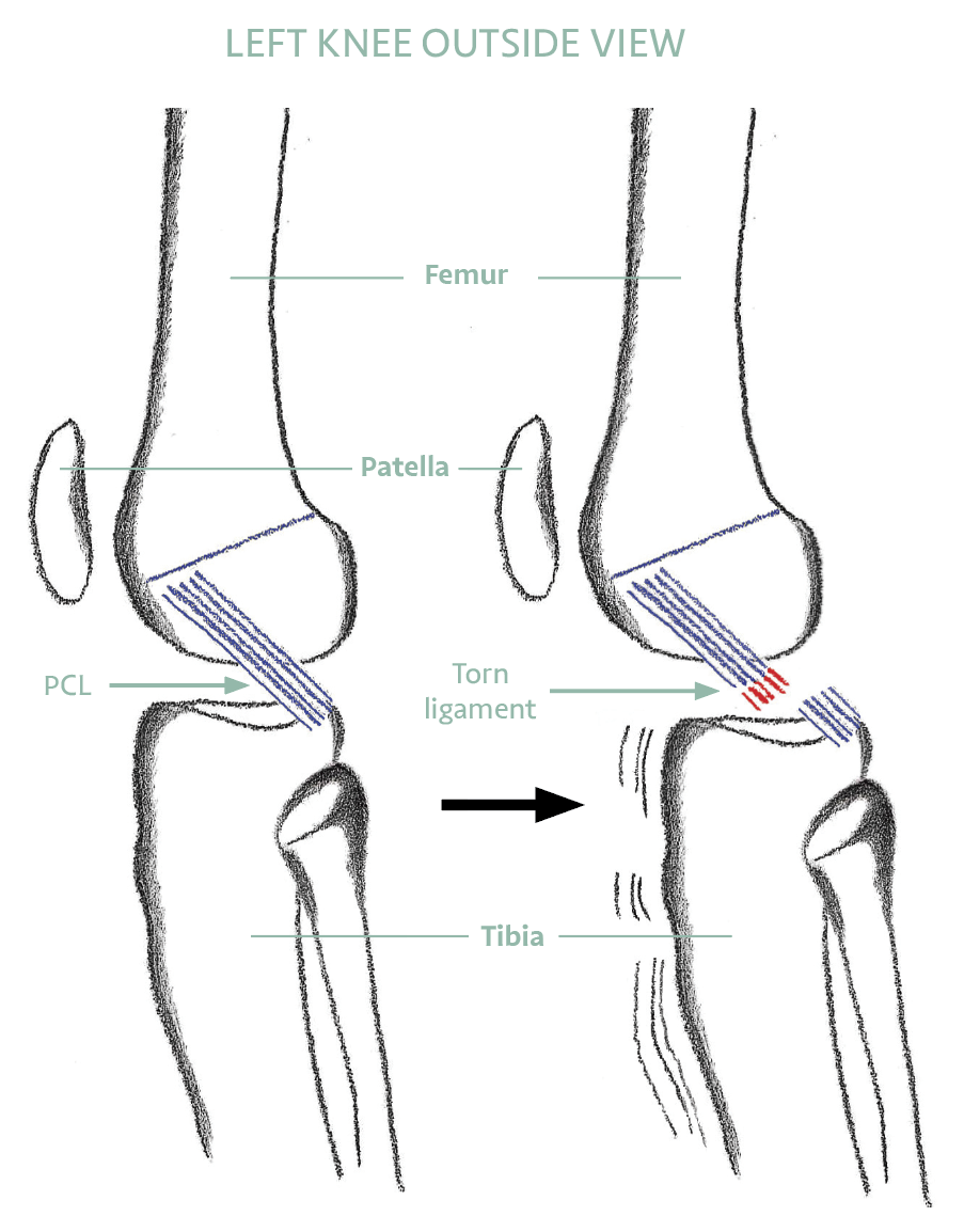
What the PCL Does

The PCL is the primary restraint against the tibia sliding backwards relative to the femur. It is approximately twice as strong as the ACL and has a better blood supply, which contributes to its superior capacity to heal without surgical intervention.

PCL injuries typically occur from a direct blow to the front of a bent knee — a dashboard injury in a car accident, a fall onto a bent knee with the foot plantar-flexed, or a direct tackle in sport. They can also occur with severe hyperextension.

Isolated PCL injuries often go undiagnosed because the initial swelling is less dramatic than an ACL injury. A posterior sag of the tibia — visible when the knee is bent at 90 degrees — is the classic clinical sign.


HOW IT WORKS WITH OTHERS

More Than One Job — and Rarely Alone

Preventing the shin from sliding backwards is the PCL’s main job, but it is not its only one. The PCL also provides quieter, secondary restraint against the knee twisting outwards and against side-to-side opening — particularly when the knee is close to straight. In the language clinicians use, it is the primary restraint to backward movement and a secondary restraint in several other directions: a genuine multi-tasker.

Just as important, the PCL rarely acts alone. The task of resisting backward movement is shared with neighbouring structures, including the meniscofemoral ligaments described in the research below, and the PCL works closely with the posterolateral corner on the outer back of the knee (Destination 9). When the PCL and the posterolateral corner are injured together, backward and rotational looseness increases far more than with a PCL injury alone. That is why a thorough assessment never stops at the PCL — it always checks the corner too, because getting the whole picture right is what makes rehabilitation succeed.

TREATMENT

Most PCL Injuries Heal Without Surgery

Rehabilitation and muscle conditioning are the cornerstone of PCL management. Quadriceps strengthening is the priority — a strong quadriceps mechanism actively prevents the tibia from sagging backwards, compensating for the damaged ligament. Hamstring exercises should be avoided early in rehabilitation as they pull the tibia posteriorly and can stress the healing tissue.

Bracing in the early stages provides protection and confidence. Most patients with isolated PCL injuries return to full activity within 3 to 6 months depending on the grade of injury.


NON-OPERATIVE MANAGEMENT

  • Most isolated PCL injuries managed without surgery
  • Bracing in early stages for protection and comfort
  • Quadriceps strengthening is the rehabilitation priority
  • Hamstring exercises avoided early — they increase posterior tibial stress
  • Cardiovascular conditioning maintained throughout
  • Return to sport: 3 to 6 months depending on grade

WHEN SURGERY MAY BE NEEDED

  • Persistent instability despite full rehabilitation
  • Multiple ligament injury — PCL with ACL or PLC
  • Bony avulsion fracture requiring reattachment
  • High-demand athlete with a Grade 3 injury

‘PCL surgery is only required in persistent instability or multiple ligament injuries. Most PCL injuries respond very well to a focused rehabilitation programme.’


YOUR SELF-CHECK: Does your knee feel unstable going downstairs, on slopes, or when decelerating suddenly? This posterior instability is the hallmark of significant PCL injury and warrants clinical assessment. A quiet ache at the back of the knee that worsens with activity can also be a PCL sign.


EVIDENCE BASE

What the Research Shows

REFERENCES — CLINICAL REFERENCES

  1. Shelbourne et al. (Am J Sports Med, 1999) — Long-term outcomes of non-operatively treated PCL injuries show good results in most patients with isolated tears.
  2. Swinford ST, LaPrade R, Engebretsen L, Cohen M, Safran M (2020) — Biomechanics and physical examination of the posteromedial and posterolateral knee: state of the art. J ISAKOS. Source for the PCL’s secondary restraint roles and its combined function with the posterolateral corner.
  3. LaPrade et al. (JBJS, 2015) — PCL reconstruction: indications, surgical techniques, and outcomes.
  4. Gupte CM, Bull AMJ, Thomas RD, Amis AA (2003) — The meniscofemoral ligaments: secondary restraints to the posterior drawer. Journal of Bone and Joint Surgery Br 85(5):765–773. Mr Gupté’s PhD research demonstrating that the meniscofemoral ligaments assist the PCL in restraining posterior tibial draw — directly relevant to understanding PCL biomechanics and why their preservation matters during PCL surgery.
  5. Amis AA, Gupte CM, Bull AM, Edwards A (2006) — Anatomy of the posterior cruciate ligament and the meniscofemoral ligaments. Knee Surgery, Sports Traumatology, Arthroscopy 14:257–263. Co-authored definitive anatomical study of the PCL and related structures.
  6. Amadi HO, Gupte CM et al. (2008) — A biomechanical study of the meniscofemoral ligaments and their contribution to contact pressure reduction in the knee. Knee Surgery, Sports Traumatology, Arthroscopy 16:1004–1008. Shows that retention of the meniscofemoral ligaments is advisable in PCL surgery to protect the lateral compartment.
  7. Gupte CM, Shaerf DA, Sandison A, Bull AMJ, Amis AA (2014) — Neural structures within human meniscofemoral ligaments: a cadaveric study. Anatomy Research International. Demonstrates proprioceptive neural elements in the ligaments adjacent to the PCL, with implications for functional rehabilitation after PCL injury.
  8. Gupte CM, Smith A, McDermott ID, Bull AMJ, Thomas RD, Amis AA (2002) — Meniscofemoral ligaments revisited: anatomical study, age correlation and clinical implications. Journal of Bone and Joint Surgery Br 84(6):846–851. The foundational cadaveric anatomy paper establishing the anatomy of the structures that assist the PCL.
  9. Gupte CM, Smith A, Jamieson N, Bull AMJ, Thomas RD, Amis AA (2002) — Meniscofemoral ligaments: structural and material properties. Journal of Biomechanics 35:1623–1629. Establishes the load-bearing properties of the meniscofemoral ligaments, which act in tandem with the PCL to resist posterior tibial draw.

PCL Assessment

Get a clear diagnosis and rehabilitation plan from the SportsHealing team.